Category: Tropical papers

How does cornea by onchocerciasis look like?

“The characteristic facial expression of the onchocerciasis patients is also based on the interstitial keratitis, which prefers a horizontal axis and creates a vertical positioned ovular bow in the ribbon shaped degeneration of the cornea”. (c) Dr. Guido Kluxen’s excellent reasearch book “Dr. Jean Hissette’s Research Expeditions to Elucidate River Blindness”, 2011

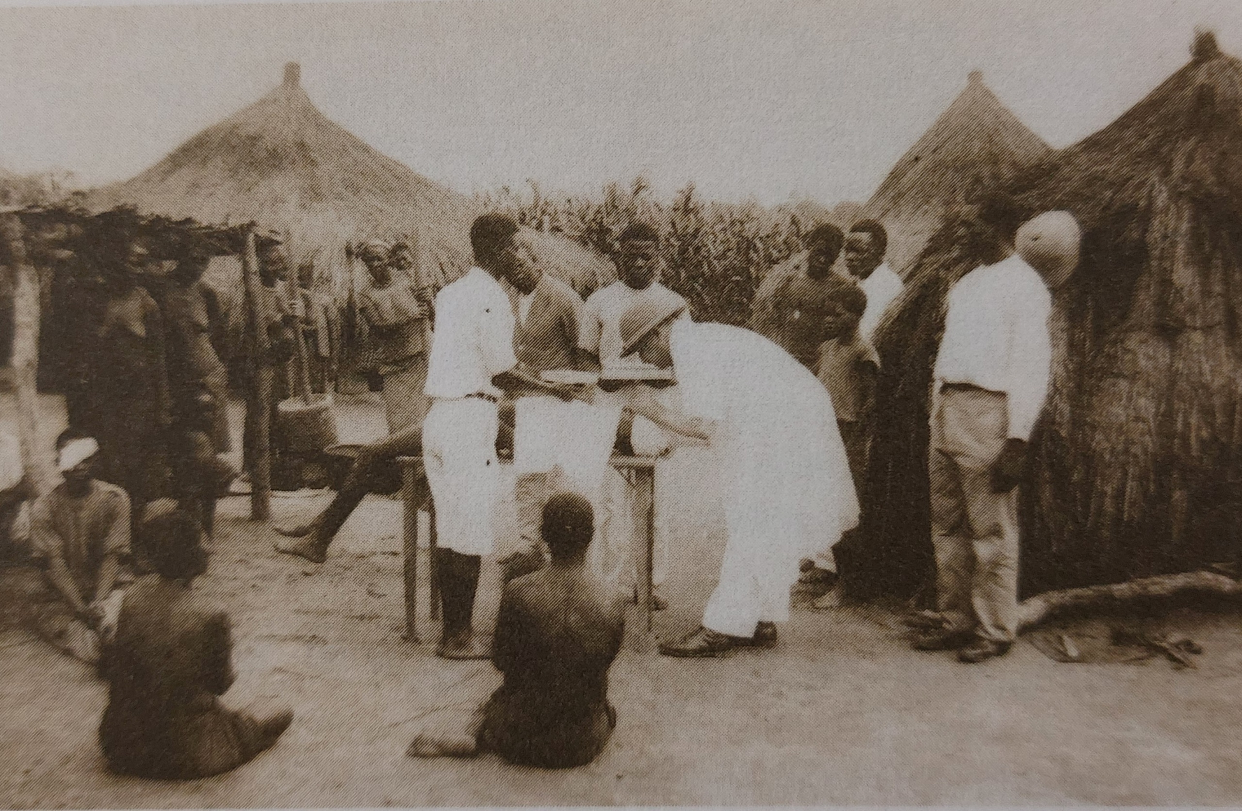
I think I had never seen onchocerciasis patients in Zambia. Although onchocerciasis is not endemic there, one sees daily unclear corneal opacities as a result of various keratitides or uveitides. Some patients travel from Angola, Namibia and Congo. The latter should still be endemic… So I decided to google these corneas, as the description is not too descriptive.

Keratitis semilunaris, sclerosing keratitis by onchocerciasis, (c) https://www.researchgate.net/figure/Keratitis-semilunaris-a-form-of-the-band-shaped-keratopathy-by-courtesy-of-HjTrojan_fig4_224830713

Cataract surgery outreach, Congo, 1930s

Dr. Jean Hissette’s cataract surgery in Kasai in the Belgian Congo, 1930

Photo from Dr. Guido Kluxen’s excellent reasearch book “Dr. Jean Hissette’s Research Expeditions to Elucidate River Blindness”, 2011

Interestingly, Dr. Hissette performed his cataract OPs using large conjunctival peritomies and additionally – iridectomies. Many surgeries were complicated with synechias, which were typicall for onchocerciasis induced uveitis.

I found interesting as well, how the doctor treated assistance during these OPs: “I did the surgeries without assistance, as I believe that if you do not have adequate assistance, it is better to operate without any”. May be often the case in the tropical setup.

Dr. Jean Hissette’s cataract surgery in Kasai in the Belgian Congo, 1930 (Photo from Dr. Guido Kluxen’s excellent reasearch book “Dr. Jean Hissette’s Research Expeditions to Elucidate River Blindness”, 2011)

Fungal keratitis – neglected tropical disease

https://pubmed.ncbi.nlm.nih.gov/36294612/

Not every fornix-OSSN is to be exenterated

In our Setup in Zambia one rule which we used to have was: as soon as OSSN involves a caruncle or conjunctival fornices, the eye (no matter how good it could see) should be planned to exenteration or at least extended enucleation). I used to not to question this practice in the abscence of MRI at hand.

However, fellows from Duisburg (Germany) proved that an excision of forniceal OSSN can still be good compensated with buccal or amniotic mucous membrames. And recurrencies of ~30%. Which is actually not bad considering the mutilating alternative. A food for thought. https://pubmed.ncbi.nlm.nih.gov/35940212/