Tag: retinoblastoma

Role of CT in Diagnosis and Monitoring of Retinoblastoma in Africa

Authors from the Western Africa have concluded that CT-Scan plays an important role by retinoblastoma cases in developing countries, despite its well-known carcinogenic ability by this cohorta of patients. Lack of MRI in the most remote areas justifies the use of CT, according to authors. Thus they were able to reveal trilateral retinoblastoma cases and extraocular extension corresponding good with histhopathological evaluation of specimens.

J Fr Ophtalmol. 2019 Dec;42(10):1085-1089. doi: 10.1016/j.jfo.2019.06.003. Epub 2019 Sep 24.
[Role of CT in Diagnosis and Monitoring of Retinoblastoma in Senegal]
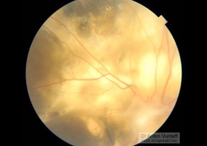
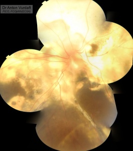
A S Sow 1 , J M M Ndiaye 2 , A M Ka 3 , D G T Sacramento 2 , H Kane 2 , M Nguer 2 , J P Diagne 3 , A M Wane 2 , E A Ba 2 , P A Ndoye Roth 2 , P A Ndiaye 3

Introduction: Retinoblastoma is a malignant tumor of neuroepithelial origin, developed from young retinal cells, occurring in infants and young children. The goal of the study was to assess the role of CT in the diagnosis of retinoblastoma at the Aristide le Dantec Hospital in Dakar.

Patients and methods: This is an 11-year retrospective study of 160 patient records in the ophthalmology department and pediatric oncology unit of the same hospital.

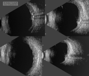
Results: One hundred and sixty (160) patients were recruited. The male:female ratio was 1.05. The mean age was 32.19 months. Leukocoria was the most common clinical sign, occurring in 105 cases (65.62 %). The retinoblastoma was intraocular in 97 cases (60.62 %). We saw 122 (76.25 %) unilateral and 3 (1.87 %) trilateral presentations. CT was performed in 150 children with 110 cases (73.33 %) of calcifications ; 62 cases (41.33 %) of optic nerve invasion ; 24 cases (16 %) of extraocular muscle invasion and 18 cases (12 %) endocranial extension. Associated tumors were found on CT: 2 cases of suprasellar mass and 1 case of pineoblastoma. In patients whose specimens were analyzed, histology showed 48.15 % optic nerve invasion, consistent with the CT findings.

Discussion: CT has an important role in the diagnosis of retinoblastoma, despite its recent contraindication in bilateral and unilateral multinodular forms.

Conclusion: CT is a good alternative to MRI in the diagnosis of retinoblastoma in developing countries with limited technological resources.

Retinoblastoma, group E

In English

In EnglishCase of April, 2016. This was a 4 yo boy, in whose eye mother had noticed white stuff one week before presentation. Video shows the status of the posterior segment via biomicroscopy. Gross white retinal tumor. Far vitreous tumor seeds. Concurrent wide retinal detachment. B-scan showed intraocular tumor with more than 10 mm base, with vitreous seeds and diffuse calcifications within the tumor.
This retinoblastoma grade is “E” (international retinoblastoma classification, the tumor occupies more than half of the eyeball). The wide spread treatment at the time for group E was (and probably still is) enucleation. Intravenous chemotherapy is mainly indicated in case “high-risk” tumor was confirmed via pathology. However, intraarterial chemotherapy was shown to be effective in eye salvage in some cases (Evidence for Stage-based Chemotherapy, 2015).
In African setup, in Kenya, Tansania, Zambia and other countries of COECSA region, the protocol for retinoblastoma treatment exists. However, the threshold for enucleation is very low, due to the known statistics: in Africa, only 30% of children with retinoblastoma survive. At the same time, it is 97% in the Western world.
The eye was enucleated, and the tumor was not proved to be “high risk”, i.e. no intrusion beyound the eye wall or into the optic nerve section.
The wide spread practice in the World is to implant orbital prosthesis simultaneously after enucleation. This can be not the case in rural African setup, where no skills or prostheses are often available. Postponed eye posthesis is also possible, with worse esthetic and prosthesis function prognosis due to fast developing asymmetry in kids’ orbits and fast conjunctiva contraction in empty socket.

in Russian / по-русскиСлучай апреля 2016 г.

Ретинобластома Группа “Е”. Мальчик 4 лет. Мама заметила белый зрачок неделю назад. Картина при биомикроскопии – на видео. Биомикроскопия: Обширная белая опухоль сетчатки. Отсевы далеко в стекловидное тело. Массивная отслойка сетчатки. По результатм ультразвукового исследования: массивная внутриглазная опухоль с оcнованием более 10 мм, с дальними отсевами в стекловдное тело, с диффузными кальцификатами.

Данная ретинобластома относится к группе E по международной классификации ретнобластом. Лечение опухолей из группы Е – энуклеация. Интраартериальная химиотерапия улучшает прогноз при применении в высокорисковом глазу. Глаз был энуклеирован. Второй глаз – не вовлечён.

В заключении гистологов: внутриглазная опухоль без прорастания в зрительный нерв без вовлечения оболочек, с обширным внутренним некрозом, “маленькие круглые голубые клетки”. Выживаемость при ретинобластоме в странах Африки = 30%, на Западе – до 97%.

https://www.youtube.com/watch?v=F7oruQBDaRE

To remove or not to remove? Coats Vs Retinoblastoma

In EnglishThis a boy of 6 yo. His other eye is normal, but left eye has VA Hand movement. He does not recall well when it had become like this, but probably about 2 years back. Parents are also not very helpful with the history.

His left fundus has yellow reflex. Anterior segment is normal, and IOPs are normal. Gonioscopy reveals normal angles’ anatomy.

At the fundus I can see a disk of peculiar shape and extensive fibrosis, some silicon oil-like white bubbles in the retina, some sheathing of the inferior vessels, massive exudates. No obvious telangiectasias.

Ultrasound shows some calcifications, and no obvious exudative retinal detachment.

It doesn’t seem like retinoblastoma (more likely – Coats?), but calcifications make us worry. Should it be enucleation just because of the calcifications? Pubmed says, Coats with calcifications does exist, but this is very rare. And literature says as well, Coats is a commonest disease confused with retinoblastoma and leading to enucleation by mistake. Sometimes this can be unavoidable.

To help us establish the correct management, we are referring the boy therefore for the CT scan of the chest, Ultrasound of the abdomen and preauricular lymph-nodes, and the neck. It would also be helpful if the MRI of orbits and a brain is available before any interventions.

Enucleation with orbital implant and artificial eye is a most likely option in our opinion.

Thanks to Dr. Tatiana Ushakova for the proper investigations consultation.

Presumably Coats disease: peculiar silicon oil-like round inclusions in the retina


Presumably Coats disease

Presumably Coats disease


Presumably Coats disease

Presumably Coats disease. Calcifications on B-scan.