![]() This a boy of 6 yo. His other eye is normal, but left eye has VA Hand movement. He does not recall well when it had become like this, but probably about 2 years back. Parents are also not very helpful with the history.
This a boy of 6 yo. His other eye is normal, but left eye has VA Hand movement. He does not recall well when it had become like this, but probably about 2 years back. Parents are also not very helpful with the history.
His left fundus has yellow reflex. Anterior segment is normal, and IOPs are normal. Gonioscopy reveals normal angles’ anatomy.
At the fundus I can see a disk of peculiar shape and extensive fibrosis, some silicon oil-like white bubbles in the retina, some sheathing of the inferior vessels, massive exudates. No obvious telangiectasias.
Ultrasound shows some calcifications, and no obvious exudative retinal detachment.
It doesn’t seem like retinoblastoma (more likely – Coats?), but calcifications make us worry. Should it be enucleation just because of the calcifications? Pubmed says, Coats with calcifications does exist, but this is very rare. And literature says as well, Coats is a commonest disease confused with retinoblastoma and leading to enucleation by mistake. Sometimes this can be unavoidable.
To help us establish the correct management, we are referring the boy therefore for the CT scan of the chest, Ultrasound of the abdomen and preauricular lymph-nodes, and the neck. It would also be helpful if the MRI of orbits and a brain is available before any interventions.
Enucleation with orbital implant and artificial eye is a most likely option in our opinion.
Thanks to Dr. Tatiana Ushakova for the proper investigations consultation.
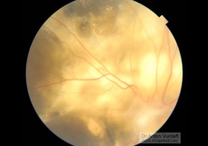
Presumably Coats disease: peculiar silicon oil-like round inclusions in the retina
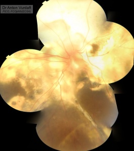
Presumably Coats disease
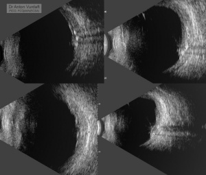
Presumably Coats disease. Calcifications on B-scan.