Tag: uveitis

Primary Vitreoretinal Lymphoma in HIV Infection.

Steffen J1, Coupland SE2,3, Smith JR4.
Author information:
1. Division of Ophthalmology, University of Cape Town, Cape Town, South Africa.
2. Institute of Translational Medicine, University of Liverpool, Liverpool, UK.
3. Liverpool Clinical Laboratories, Liverpool University Hospital NHS Foundation Trust, Liverpool, UK.
4. Eye and Vision Health, Flinders University, Adelaide, Australia.

Abstract

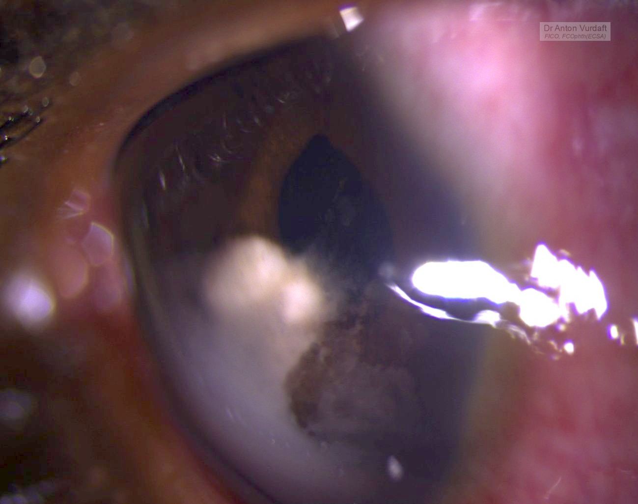
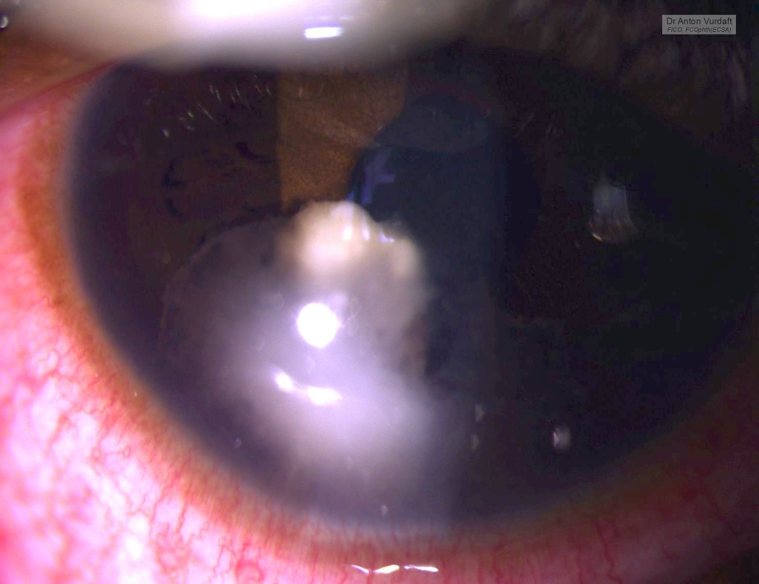
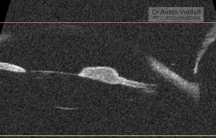
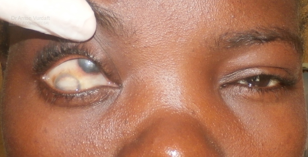
Purpose: To describe the epidemiology, clinical characteristics, diagnosis and treatment of human immunodeficiency virus (HIV)-related primary vitreoretinal lymphoma (PVRL).Methods: Narrative literature review.Results: HIV-related PVRL occurs in persons who are relatively young and generally have very low CD4+ T-cell counts. Vitritis with subretinal or sub-retinal pigment epithelial infiltrates is typical. Vitreous cytology remains the gold standard for diagnosis, supplemented by flow cytometry and genetic analyses of tumor cells, and measurement of aqueous or vitreous interleukin-10 levels. Concurrent brain involvement also may establish the diagnosis. Treatment includes antiretroviral therapy (ART), systemic chemotherapy (usually methotrexate-based) and local ocular treatment (intravitreal methotrexate, intravitreal rituximab, external beam radiotherapy). Systemic chemotherapy is of uncertain value for PVRL without other central nervous system involvement. Prognosis is poor, but has improved significantly compared to the pre-ART era. Conclusions: Ophthalmologists should consider the diagnosis of PVRL in HIV-positive individuals who present with intermediate or posterior uveitis.

PMID: 32453669

Anterior chamber and iris granuloma in newly diagnosed HIV+ kid

This was a case of Mai 2016. Eleven y.o. boy came to our outpatient department with the father complaining of “growth” in one of boy’s eyes, that was allegedly present already for 2 weeks. Visual acuity 6/9 and there were no pains except photophobia.  On examination – solid feathery cream-colored mass in the AC, adherant to the iris. Yellowish behind. No anterior chamber cells, no flare, absolutely quiet eye. Upon the HIV-test she was confirmed newly positive.

presumed aspergillosis iris-AC granuloma

presumed aspergillosis iris-AC granuloma

She was started on steroid and cycloplegic topical eye drops and NSAID tablets, and in 5 days the exudate was gone. But the iris cyst remained in place. There was no vitreous or retinal involvement, and the IOP was OK.

presumed aspergillosis iris-AC granuloma after tx, note torn posterior synechiae

presumed aspergillosis iris-AC granuloma after tx: UBM-scan

Differential diagnosis: intraocular medulloepithelioma (must be congenital, but looks similar, although originating mostly from the angle or ciliary body, not the iris), intraocular lymphoma (looks simmilar, but much more scary!), tuberculoma, leproma, nonpigment melanoma or primary iris tumors, toxoplasmosis gumma, metastases in the iris (lungs, intestine, kidneys), fungi!

Other useful resources  to review iris cysts:

eyewiki.aao.org/Iris_Cysts
bjo.bmj.com/content/59/5/276.full.pdf
https://www.archivesofpathology.org/doi/pdf/10.5858/arpa.2010-0669-RS
http://www.ncbi.nlm.nih.gov/pmc/articles/PMC3872564/

Literature search at that time in 2016 showed one paper from 2009 by Jain V with a similar case, Aspergillus iris granuloma: a case report with review of literature, claiming that to 2009 only three cases of aspergillosis of the anterior segment of the eye had been ever published. Here is a picture of their case:

Jain V, Aspergillus iris granuloma: a case report with review of literature. 2009

Our patient was left on the same topical treatment further in attempt to conquer the cyst and lost to follow-up. Unfortunately or fortunately, no iris-biopsy was performed that time, and therefore I had no microbiological evidence of fungi. One must also consider, that no natamycin was readily available topically in Western Province of Zambia that time, and in most of the times one must rely on antifungal activity of povidone-iodine eye drops (which are good and readily available hand made, by the way). One must strongly consider the lungs screening (CXR or CT) in immunocompromised patients, to exclude the primary source of hematogenous dissemination to
the brain or meninges. The immune status must be supported of course (f.e. HAART).

I thank my peer-colleagues from Terra-Ophthalmica for the kind help with the differential diagnosis and additional ideas for this case.

Pupula duplex

In EnglishThis is a young lady with a case of old scleral malacia and uveal staphyloma. This staphyloma actually looks very much like a typical advanced “anterior staphyloma”, which usually originates at the cornea site. She also has phthitic changes of her real cornea and anterior chamber, which is positioned infero-laterally.

In other words, such or similar cases could have been the source for the very popular rumor/myth in ophthalmology, that there were cases of double corneas+irises+pupillae. They call it “pupula duplex” (sic) and photoshop pictures are all over the google. I have read people also considered this as “devil eye”. Very much possible:)

In reality, however, this lady has all the indications for enucleation, histopathology and implant/prosthesis with a tumor suspect via B-scan. Her current condition is there since 2015.

That is what potential Pupula duplex case could look like when it supposedly was described in older literature, if true at all. “Real” cornea is lower, “pseudo” cornea is upper.