Tag: HIV

Primary Vitreoretinal Lymphoma in HIV Infection.

Steffen J1, Coupland SE2,3, Smith JR4.
Author information:
1. Division of Ophthalmology, University of Cape Town, Cape Town, South Africa.
2. Institute of Translational Medicine, University of Liverpool, Liverpool, UK.
3. Liverpool Clinical Laboratories, Liverpool University Hospital NHS Foundation Trust, Liverpool, UK.
4. Eye and Vision Health, Flinders University, Adelaide, Australia.

Abstract

Purpose: To describe the epidemiology, clinical characteristics, diagnosis and treatment of human immunodeficiency virus (HIV)-related primary vitreoretinal lymphoma (PVRL).Methods: Narrative literature review.Results: HIV-related PVRL occurs in persons who are relatively young and generally have very low CD4+ T-cell counts. Vitritis with subretinal or sub-retinal pigment epithelial infiltrates is typical. Vitreous cytology remains the gold standard for diagnosis, supplemented by flow cytometry and genetic analyses of tumor cells, and measurement of aqueous or vitreous interleukin-10 levels. Concurrent brain involvement also may establish the diagnosis. Treatment includes antiretroviral therapy (ART), systemic chemotherapy (usually methotrexate-based) and local ocular treatment (intravitreal methotrexate, intravitreal rituximab, external beam radiotherapy). Systemic chemotherapy is of uncertain value for PVRL without other central nervous system involvement. Prognosis is poor, but has improved significantly compared to the pre-ART era. Conclusions: Ophthalmologists should consider the diagnosis of PVRL in HIV-positive individuals who present with intermediate or posterior uveitis.

PMID: 32453669

Anterior chamber and iris granuloma in newly diagnosed HIV+ kid

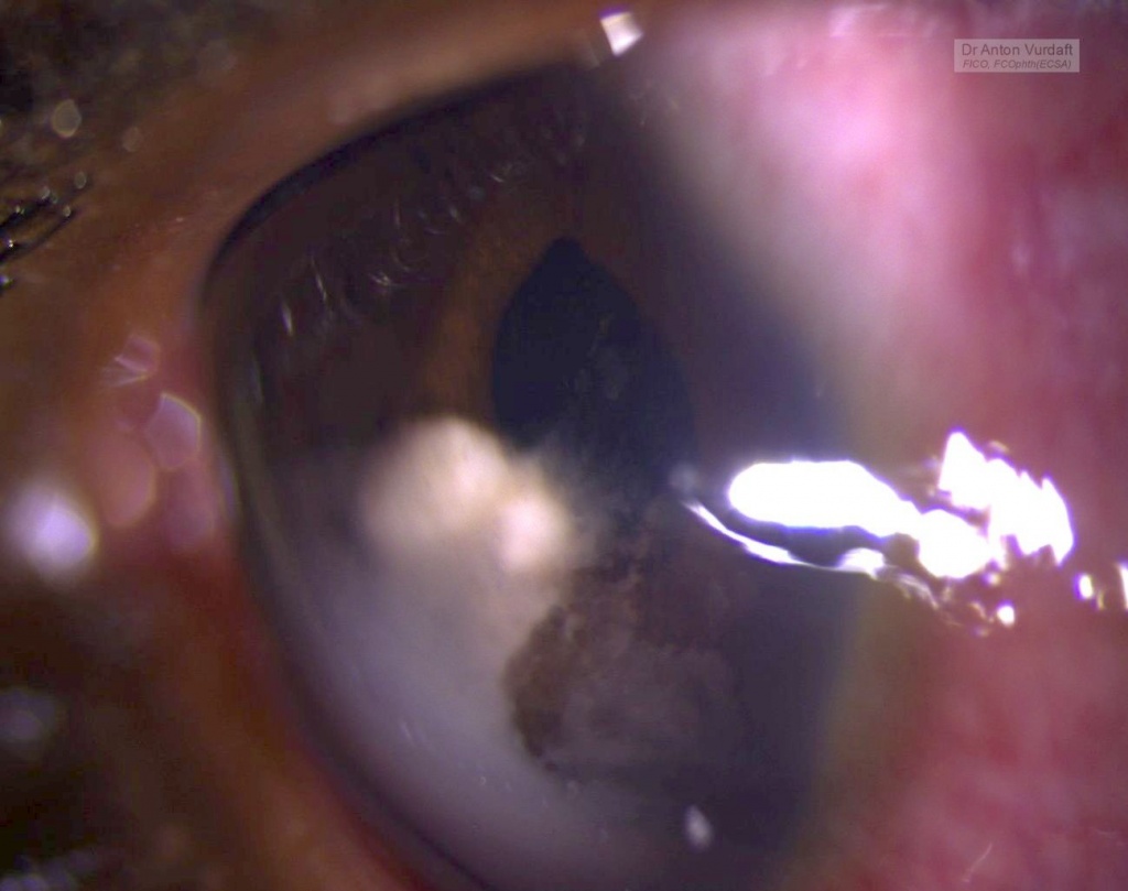
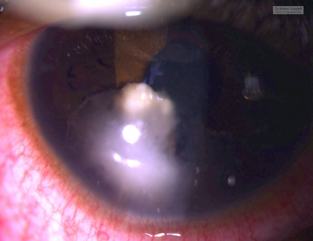
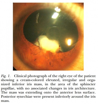
This was a case of Mai 2016. Eleven y.o. boy came to our outpatient department with the father complaining of “growth” in one of boy’s eyes, that was allegedly present already for 2 weeks. Visual acuity 6/9 and there were no pains except photophobia.  On examination – solid feathery cream-colored mass in the AC, adherant to the iris. Yellowish behind. No anterior chamber cells, no flare, absolutely quiet eye. Upon the HIV-test she was confirmed newly positive.

presumed aspergillosis iris-AC granuloma

presumed aspergillosis iris-AC granuloma

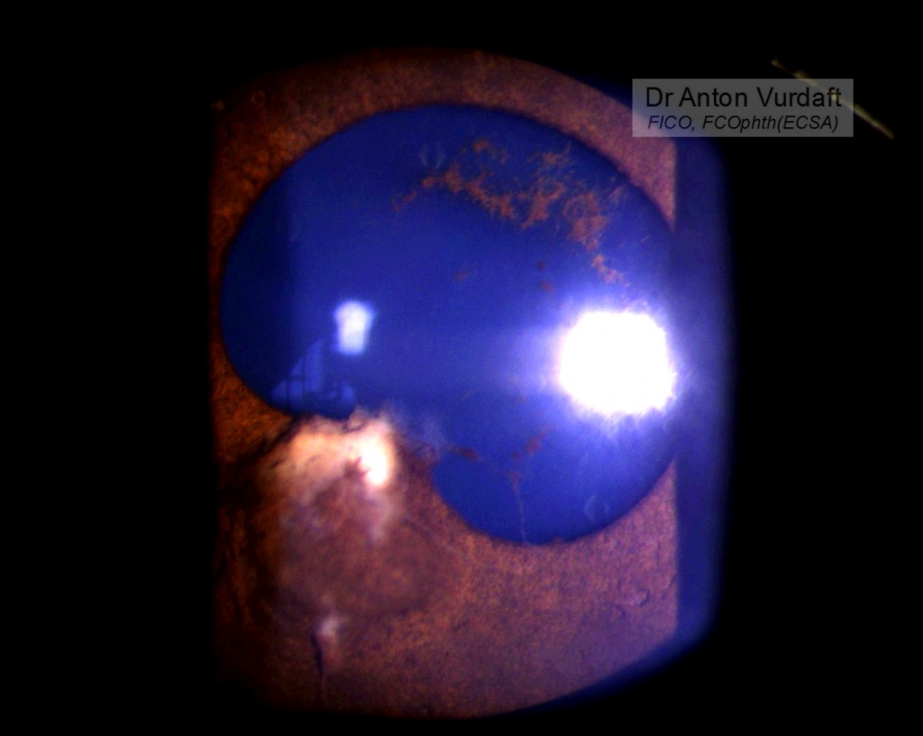
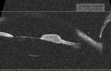
She was started on steroid and cycloplegic topical eye drops and NSAID tablets, and in 5 days the exudate was gone. But the iris cyst remained in place. There was no vitreous or retinal involvement, and the IOP was OK.

presumed aspergillosis iris-AC granuloma after tx, note torn posterior synechiae

presumed aspergillosis iris-AC granuloma after tx: UBM-scan

Differential diagnosis: intraocular medulloepithelioma (must be congenital, but looks similar, although originating mostly from the angle or ciliary body, not the iris), intraocular lymphoma (looks simmilar, but much more scary!), tuberculoma, leproma, nonpigment melanoma or primary iris tumors, toxoplasmosis gumma, metastases in the iris (lungs, intestine, kidneys), fungi!

Other useful resources  to review iris cysts:

eyewiki.aao.org/Iris_Cysts
bjo.bmj.com/content/59/5/276.full.pdf
https://www.archivesofpathology.org/doi/pdf/10.5858/arpa.2010-0669-RS
http://www.ncbi.nlm.nih.gov/pmc/articles/PMC3872564/

Literature search at that time in 2016 showed one paper from 2009 by Jain V with a similar case, Aspergillus iris granuloma: a case report with review of literature, claiming that to 2009 only three cases of aspergillosis of the anterior segment of the eye had been ever published. Here is a picture of their case:

Jain V, Aspergillus iris granuloma: a case report with review of literature. 2009

Our patient was left on the same topical treatment further in attempt to conquer the cyst and lost to follow-up. Unfortunately or fortunately, no iris-biopsy was performed that time, and therefore I had no microbiological evidence of fungi. One must also consider, that no natamycin was readily available topically in Western Province of Zambia that time, and in most of the times one must rely on antifungal activity of povidone-iodine eye drops (which are good and readily available hand made, by the way). One must strongly consider the lungs screening (CXR or CT) in immunocompromised patients, to exclude the primary source of hematogenous dissemination to
the brain or meninges. The immune status must be supported of course (f.e. HAART).

I thank my peer-colleagues from Terra-Ophthalmica for the kind help with the differential diagnosis and additional ideas for this case.

COVID-19, a thought of the day

It’s sad to realize how ignorant a western community was and is when it comes to ebola, HIV, TB and malaria African epidemics, eliminating annually the whole layers of population. And how suddenly important epidemics become when the seek persons walk around the BBC and CNN headquarters. Define “hypocrisy“…

What do you know about HIV transmission in ophthalmology surgery?

In EnglishThe following information is essential for those colleagues-ophthalmologists travelling abroad into the tropics, where the rate of HIV may reach up to 10-20% of a population or even more, depanding on the area. An especially for the oculoplastic colleagues. And here’s why:

Rate of gloves punctures during ophthalmic procedures reaches 21.8%!
Cataract surgeons experience only 11.9% of punctures.
Oculoplastic procedures carry the biggest risk for gloves perforation: 41.67%!
Risks increase rapidly for a longer procedures.

(Glove Perforation – Miller & Apt – Arch Ophthalmol – Vol 111, Feb 1993)

When double gloved – punctures will frequently be limited by the outer pair of gloves. Two layers may also reduce the amount of blood actually got into the skin with the sharps.

So, you have had an incident. You’ve pricked yourself with a suture needle or any other sharp during the surgery. What are your steps?

1) Stop the surgery immediately.
2) Wash your hands with soap. The CDC guidelines wouldn’t advise spirit or povidon iodine wiping, but the temptation is highest.
3) Scrub in and complete the case.
4) This is definitely the end of your operating day today, because:
5) You need to get the status of HIV of the patient as fast as possible. Ideally you have got the test done for all your patients before the surgeries.
6) The patient found positive (by fresh test or via previous history).
7) You test yourself as well (as per protocol, the post-exposure prophylaxis is not indicated if the one is already HIV-positive).
8) You are negative. Check your liver function tests and creatinine.
9) Initiate the postexposure HIV prophilactic therapy (“PEP”). This is a set of 3-4 HAART drugs. Please, take the best PEP combination with yourself to your trips, regardless of the stocks available on sites (you never know their quality!).
10) This is for 28 days, and not without side effects, such as transitory jaundice, diarrheoa/vomitting (depending on therapy applied) and rash/pruritus.
10) Keep monitoring your side effects with the HIV-specialist. Monitor liver function tests PRN.

What works for you in this situation:

1) The risk for HIV transmission is very low by default – about 0,3% (that is good)
2) You used two pairs of gloves (that is clever in these regions!)
3) Needles from sutures carry less risk for transmission than hollow needles for injections
4) The patient might be on HAART already, this reduces his viral load to less than 1700 copies per ml (and that is awesome, risks go even lower)
5) The PEP, which you have started within two hours (24-48 hours at most!, the more – the worse), reduces the remaining lowest chances to close to zero.

What works against you:
1) High HIV viral load in the source patient (more than 1700 copies per ml increase chances for transmission 16 times, according to some papers).
2) Late initiation of PEP (studies on monkies show, that late start – after 48 hours – is almost useless, as it allows more than 50% “lucky” guys be infected still).

P.S. I am not talking about the situation, when the patient is found negative, though it can be a very tricky question. Remember, there are window periods for the HIV to become detectable by routine express tests.

If there is even a slight level of uncertainty – you must start the PEP.

Stay safe!