Tag: HIV

Vernal-like keratoconjunctivitis in adults with HIV

An Entity, of which I was not aware…

The role of human immunodeficiency virus in the pathogenesis of vernal keratoconjunctivitis-like disease in adults: A demographic and epidemiological study
Indian J Ophthalmol. 2020 Aug;68(8):1551-1554. doi: 10.4103/ijo.IJO_1608_19.
Authors
Anine Kritzinger 1 , Anthony G Zaborowski 1 , Wilbert Sibanda 2 , Linda Visser 1
Affiliations
1 Department of Ophthalmology, University of KwaZulu-Natal, Durban, South Africa.
2 School of Mathematics, Statistics and Computer Science, University of KwaZulu-Natal, Durban, South Africa.
PMID: 32709773
PMCID: PMC7640813
DOI: 10.4103/ijo.IJO_1608_19
Free PMC article
Abstract
Purpose: The purpose was to identify and describe patients with new-onset vernal keratoconjunctivitis-like (VKC-like) disease after puberty.

Methods: The study consisted of two parts: a prospective observational descriptive study of patients with new-onset VKC-like disease, and a case-control study to determine the relationship of a CD4 count with VKC-like disease in adults, in the setting of human immunodeficiency virus (HIV). Patients were recruited between January 2016 and November 2017 from a Provincial Eye hospital, one of two large referral hospitals in KwaZulu-Natal, South Africa. Patients presenting to the eye clinic were screened and diagnosed at the Primary Eye Care Unit. Inclusion criteria: age 15 years and older with signs and symptoms of new-onset VKC-like disease. Exclusion criteria: a history of childhood atopic diseases, atopic keratoconjunctivitis and patients who declined HIV testing. Data collected included HIV status, CD4 count, antinuclear antibodies and total serum immunoglobulin E.

Results: Thirty-three patients were included; females n = 16 and males n = 17. The mean age at presentation was 32.45 ± 9.93 years, 95% CI = 28.94-35.97. Twenty-six patients (78.8%) were HIV positive, 95% CI (62-89). The proportion of HIV positive patients was statistically different from the HIV negative group, Chi-squared = 21.866, P value <0.0001. In the group of HIV positive patients, 72% were classified as immunodeficient according to their CD4 counts. An association was proven between severely immunodeficient patients and the risk of VKC-like disease (Chi-squared = 4.992, P value = 0.0255).

Conclusion: In this cohort, a statistically significant association was found between VKC-like disease in adults and an HIV positive status. This association calls for more research on the subject.

Keywords: Allergy; VKC-like disease in adults; immunocompromised; ocular manifestations of HIV; vernal keratoconjunctivitis.
Ocular surface squamous neoplasia in Zambia

In this blog one may find lots of pictures of OSSN. Finally we got some science for that!

Clinical and Pathologic Presentation of Primary Ocular Surface Tumors among Zambians
Peter Julius et al. Ocul Oncol Pathol. 2021 Mar;7(2):108-120.
doi: 10.1159/000511610. Epub 2021 Jan 21.

Authors
Peter Julius 1 , Stepfanie N Siyumbwa 1 , Phyllis Moonga 2 , Fred Maate 1 , Trevor Kaile 1 , Guobin Kang 3 , John T West 4 , Charles Wood 3 , Peter C Angeletti 3

1 Department of Pathology and Microbiology, School of Medicine, Lusaka, Zambia.
2 University Teaching Hospital, Eye Hospital, Lusaka, Zambia.
3 Nebraska Center for Virology and the School of Biological Sciences, University of Nebraska-Lincoln, Lincoln, Nebraska, USA.
4 Nebraska Center for Virology and the Department of Biochemistry, University of Nebraska-Lincoln, Lincoln, Nebraska, USA.

PMID: 33869164
PMCID: PMC8024974 (available on 2022-03-01)
DOI: 10.1159/000511610

Aim: This study aimed to characterize the clinical and pathologic presentation of ocular surface tumors (OSTs) and to more precisely differentiate the grades of ocular surface squamous neoplasia (OSSN) and benign lesions among Zambians.

Methods: Two-hundred sixty-five Zambian patients presenting with ocular surface growths, suspicious for OSSN, were recruited between November 2017 and November 2019 to a cross-sectional study to investigate their lesions. Sociodemographic data were collected, HIV infection status and vision tests were performed, and lesions were measured and documented. Lesions >2 mm in diameter were excised and sent for pathology analysis. In addition to the biopsies, tears, blood, and buccal swabs were collected. CD4+ T-cell counts were measured by flow cytometry. Lesions were classified according to the WHO guidelines. χ2 and bivariate correlations were used to analyze variable associations and strengths with phi/Cramer’s V and correlation coefficients, respectively. Binary logistics was used to adjust for covariance.

Results: In this study, 68.3% of the participants were found to be HIV positive. The most frequent diagnoses were invasive OSSN (45.3%), preinvasive OSSN (29.1%), and pterygium (22.6%). Invasive OSSN comprised keratinizing squamous cell carcinoma (SCC) (87.5%), basaloid SCC (3.3%), and spindle cell carcinoma (3.3%). Unusual carcinomas, not described previously, included hybrid SCC (5.0%) and acantholytic SCC (0.8%). Invasive OSSN had advanced tumor (T3/T4) staging (93.3%) at diagnosis. Lymphadenopathy was rare (2.3%), and metastasis was absent. Patients were mostly female (59.2%). Median age was 36 (interquartile ranges 33-41) years (ranges 18-81). Patients with invasive OSSN were more likely to present with pain (p = 0.007), redness (p = 0.034), excessive tearing (p = 0.0001), discharge (p = 0.011), bleeding (p = 0.007), reduced vision (p = 0.0001), fungating lesion (p = 0.001), and blindness (p = 0.005); location at temporal limbus (p = 0.0001), inferior limbus (p = 0.0001), or circumlimbal (p = 0.001); and extension to cornea (p = 0.006) and forniceal palpebral conjunctiva (p = 0.001). Invasive OSSN was associated with any smoking habit and alcohol consumption (p = 0.04 and 0.03, respectively). HIV positivity was strongly associated with OSSN (74.6% OSSN vs. 49.3% benign lesions; p = 0.0001; phi: 0.237 [p = 0.0001]).

Conclusion: OSTs are very common in Zambia and are strongly associated with HIV coinfection. Patients with OSSN were more likely to be HIV positive than those with pterygia. Despite the commonality of OSTs in sub-Saharan Africa, these cancers have historically been poorly characterized

Herpes Zoster Ophthalmicus in Africa

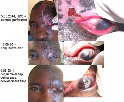
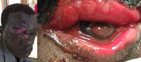
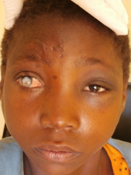
In EnglishHerpes zoster ophthalmicus involves V1 cranial nerve (n.ophthalmicus). Here are three examples of herpes zoster in 2 young men and one 7yo girl. Both men are HIV-positive (this must be an AIDS-stage). HZO is definitely second quickest germ to penetrate and destroy the cornea. Pseudomonas has no chance in comparison to those two. HIV is certainly a boost for the fulminant clinic.

in Russian / по-русскиГерпес зостер c вовлечением первой ветви из моей африканской практики. Случай у двух взрослых мужчин и у девочки 7 лет (активный у мужчин, и в исходе у девочки). Herpes zoster ophthalmicus – наверное второй по скорости разъедания роговицы после гонококковой инфекции. Синегнойная палочка определённо нервно курит в сторонке. Мужчины были однозначно ВИЧ-положительны, а это, как известно, означает стадию СПИД. И это конечно одна из основных причин для такой яркой и драматической клинической картины.

Advanced herpes zoster ophthalmicus with corneal melt and lens/uvea prolapse. HIV positive patient.

Advanced herpes zoster ophthalmicus with corneal melt and lens/uvea prolapse. HIV positive patient.

Advanced herpes zoster ophthalmicus with resultant corneal leucoma in 7yo girl.

Advanced herpes zoster ophthalmicus with resultant corneal leucoma in 7yo girl.

Advanced herpes zoster ophthalmicus with corneal melt and iris prolapse. HIV positive patient.

Advanced herpes zoster ophthalmicus with corneal melt and iris prolapse. HIV positive patient. Failed conjunctival flap adherence depicted.

Eye precancerous lesions mimic benign pyogenic granuloma

Not all pyogenic granulomas are equally safe. Some of them may comprise an elements of ocular surface squamous neoplasia, OSSN (conjunctival intraepithelial neoplasia, CIN). The paper Herwig-Carl MC, Grossniklaus HE, Müller PL, et al. Br J Ophthalmol Epub. doi:10.1136/ bjophthalmol-2018-312960 highlighted 9 such cases.

OSSN is extremely common in the areas with widespread HIV-prevalence. Human papilloma virus is responsible for this, as a typical opportunistic infection. Knowledge of the potential of OSSN to mimic pterygia and pyogenic granulomas is crucial, especially for the mid level ophthalmic personnel, as they are the primary surgeons for these conditions in many areas.

Pyogenic Granuloma Associated With Conjunctival Epithelial Neoplasia: Report of Nine Cases

Aims: To systematically describe the clinical and histopathological features of a case series of conjunctival carcinomatous lesions underlying as-and also masquerading-pyogenic granuloma.

Methods: Nine cases of conjunctival carcinomatous lesions underlying a pyogenic granuloma (which were clinically predominant) were retrospectively identified. Patients’ records were analysed for demographic data, clinical appearance and the postoperative course. Formalin-fixed paraffin-embedded specimens were routinely processed and stained with H&E and periodic acid-Schiff. Immunohistochemical stains for cytokeratin were performed in selected cases.

Results: All nine tumours were located in the conjunctiva (bulbar, tarsal, limbal conjunctiva) of patients between 44 and 80 years. The lesions exhibited clinical features of pyogenic granuloma which dominated the clinical appearance. Additional features comprised a papillomatous appearance of the adjacent conjunctiva, a more whitish aspect of the lesion and a history of squamous cell carcinoma (SCC) respectively surgery for other entities. Histopathological analysis revealed a carcinomatous lesion (conjunctival intraepithelial neoplasia or SCC) at the base of a classic pyogenic granuloma in all nine cases. Surgical removal (R0 resection) was performed. Three cases received adjuvant mitomycin C or interferon α2b treatment. Two lesions locally recurred within 2 years after initial presentation.

Conclusion: Carcinomatous lesions may be accompanied by a pyogenic granuloma which may dominate the clinical pictures. As the tumour is usually located at the base of the lesion, a complete surgical excision followed by histopathological analysis is mandatory for each lesion appearing as conjunctival pyogenic granuloma.

Keywords: conjunctival intraepithelial neoplasia; histology; pyogenic granuloma; squamous cell carcinoma; tumour.