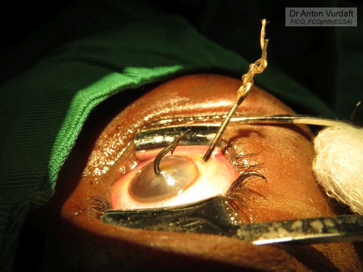
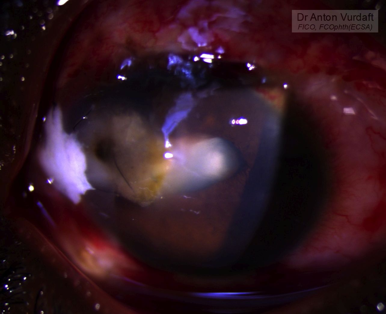
![]() EN: A case of June 2016: corneal perforation as a result of excessive corneal scrapping during pterygium excision. Although it was performed by an ophthalmic nurse, this is a rarest complication in the pterygium surgery. The surgery carries more risks, when performed without microscope, as was the case in here. The rate of complications in pterygium excisions among our ophthalmic nurses was very low. This is the only corneal perforation after such a procedure known to me in 4 years of observing nurses’ surgeries. Successfully managed with scleral autograft. One month later recurrence followed. Due to shallow anterior chamber after perforation lens had become more opaque. Recurrence and mature cataract were managed via conjunctival autograft and SICS in one setup. Video of scleral autograft is attached here.
EN: A case of June 2016: corneal perforation as a result of excessive corneal scrapping during pterygium excision. Although it was performed by an ophthalmic nurse, this is a rarest complication in the pterygium surgery. The surgery carries more risks, when performed without microscope, as was the case in here. The rate of complications in pterygium excisions among our ophthalmic nurses was very low. This is the only corneal perforation after such a procedure known to me in 4 years of observing nurses’ surgeries. Successfully managed with scleral autograft. One month later recurrence followed. Due to shallow anterior chamber after perforation lens had become more opaque. Recurrence and mature cataract were managed via conjunctival autograft and SICS in one setup. Video of scleral autograft is attached here.
![]() RU: Можно ли напортачить, удаляя птеригиум? Да, можно. Клинический случай в пример.
RU: Можно ли напортачить, удаляя птеригиум? Да, можно. Клинический случай в пример.
Классическим методом удаления головки птеригиума является “отрыв” головки механически от лимба в сторону вершины птеригиума. В случае, если такое не срабатывает, в строме под головкой остаётся много ошмётков птеригиума. Которые желательно “отшкрябать” лезвием или отшлифовать специальной машинкой. Если шкрябать слишком усердно – можно дошкрябать до передней камеры. Это особенно актуально, если птеригиум был “жирный” и если не используется микроскоп. Это крайне редкое осложнение (вижу впервые), но оказывается такое возможно.
“После сильного измельчения или спадения камеры во время перфорации катаракта его подстегнулась, и из сотых превратилась в хорошую набухшую светоощущение. Птеригиум рецидивировал, потребовал пластики. Пластика и экстракция катаракты проведены одномоментно. Вроде бы реанимировал глаз. Полёт нормальный.”
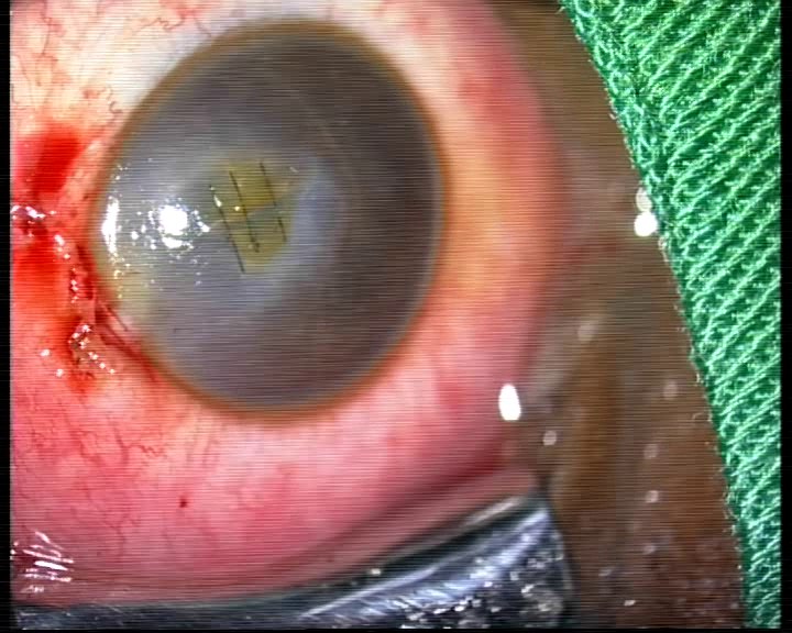
На фото – глаз пациента 65 лет, которому медсестра удалила птеригиум. Первое фото – статус после операции на третий день (с бандажной линзой). Второе – статус после закрытия дефекта (аутолоскут склеры). Далее серия фото с рецидивом и состоянием после удаления катаракты и пластики птеригиума.
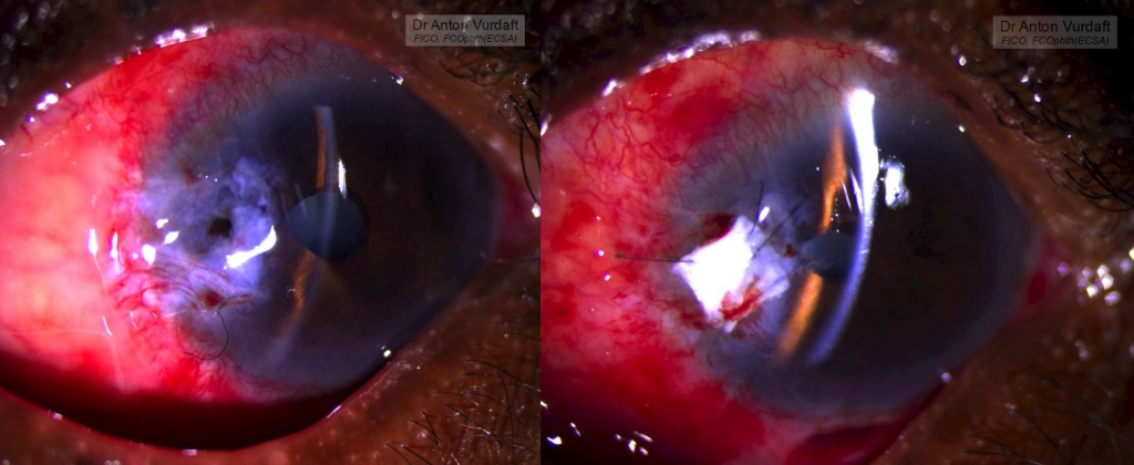
Pterygium excision caused corneal perforation
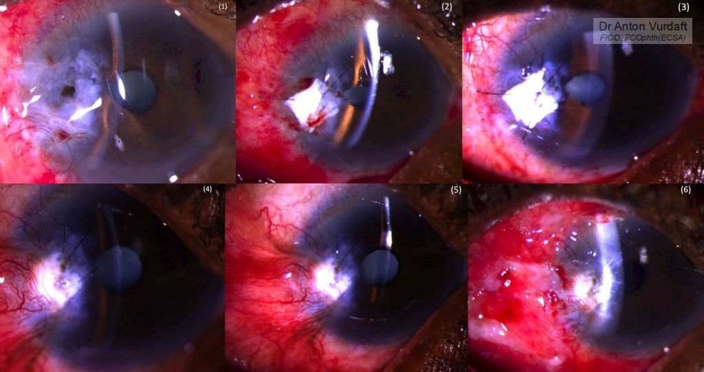
Pterygium excision caused corneal perforation. Recurrence one month later.