![]() This is a lady 20 yo, who states she had problems with right eye vision since long time back (no details, but it’s about many years). Left eye vision started deteriorating some one or two years ago. Her vision in RE is FC close to face, and LE is 6/12 (no improvement with pinhole).
This is a lady 20 yo, who states she had problems with right eye vision since long time back (no details, but it’s about many years). Left eye vision started deteriorating some one or two years ago. Her vision in RE is FC close to face, and LE is 6/12 (no improvement with pinhole).
Her current vision in RE is stable for several years, according to her, and not a recent decrease. She only acknowledges trauma to the right eye (slight punch with the tree branch) when she was very young. She also notes poor vision in dark time of the day in Left eye (since few years).
Both anterior segments are normal. Fundus exam. Right eye diffuse confluent retinoschisis with macular tear. Retina is slightly mobile throughout the posterior pole and up to periphery. No obvious funnel is seen ophthalmoscopically.
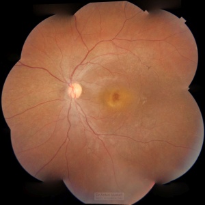
Left eye – enlarged and disturbed foveal reflex and infero-nasal periphery suspicious for retinoschisis and with lacy pattern. One bone spicule is seen at the arcade.
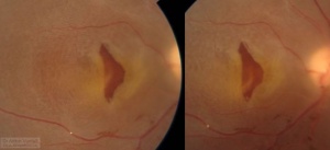
Macular tear, diffuse retinal detachment in case of juvenile maculoschisis (Stereo-pair for depth appreciation)
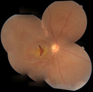
Macular tear and diffuse retinal detachment in juvenile maculoschisis. Concentric lines of retinal detachment seen throughout the fundus.
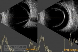
Macular tear, diffuse retinal detachment in case of juvenile maculoschisis (B-scan)
Maculoschisis (juvenile)
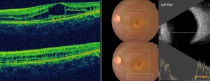
Maculoschisis (juvenile) (OCT and B-scan)
Left Eye OCT shows high foveal cysts, impending macular hole, with maculoschisis at the level of inner nuclear layer spreading far into macula.
B-scan of RE reveals funnel-shaped detachment of retina. LE shows flat schisis in macula.
Diagnosis for Right Eye: funnel shaped retinal detachment (as a complication of diffuse retinoschisis with macular tear. X-linked inheritance is unlikely (consanguinity not confirmed). Left eye impending macular hole, diffuse macular retinoschisis, peripheral retinoschisis. X-linked?
The patient was consulted with few retinal fellows through teleconsultations (Orbis Cybersight and my fellow colleagues), and current decision is to put her on topical dorzolamide TDS for both eyes. However, right eye may benefit from retinal surgery, and she was referred.
My main source for this case: Lawrence Yannuzzi “The Retinal Atlas”, 2nd Ed.