Tag: ptosis

Congenital ptosis + polypropelene sutures: hah?

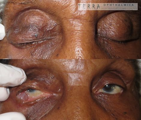
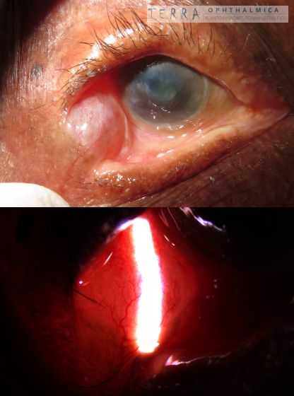
A retrospective study from Mali has shown, that in kids with congenital ptosia, in a low-resource setup the polypropelene frontalis sling could still be an option. Although the ideal material for frontalis sling is fascia lata in adults and silicone slings in kids, sutures could be an option, where those are unavailable. Authors claim ~85% success rate in 4-25 months.

I personally used this technique once in an adult lady with blepharoptosis-phimosis-canthus-inversus syndrome, with good result in a short-term. My pediatric colleague from Zambia claims also, that even with the suture cutting through, it manages meantime to create the scar, and thus to hang the lid anyway.

Source: https://www.ncbi.nlm.nih.gov/pubmed/31858999

In our study, the surgical result (prior to correction of recurrences) was satisfactory in 81.82 % of cases, with a recurrence rate of 13.64 %. The mean follow-up was 14 months, ranging from 4 to 25 months.

Dacryops (simple lacrimal gland cyst)

In English

In EnglishA case of December 2015.
Lady 75yo, presented with cyst in lateral canthal area of right eye. According to the patient, the size is dynamic, the lesion is present since 2010. She managed at least once pricking the cyst herself. She has also a history of bilateral trachoma (not treated). Trachoma is a known risk factor for lacrimal ducts scarring, which triggers so called “dacryops” – a simple cyst of lacrimal gland. The lesion is shown at the photo together with canthus involvement, corneal pannus, opacity and cataract.
DDx for etiology of dacryops nowadays is dacryoadenitis and scarring conditions of conjunctiva apart from trachoma (Stevens-Johnson, cicatrizing pemphigoid, chemical burns). In trachoma-endemic regions trachoma is the main reason for dacryops.
Marsupialization of cyst or its complete excision is indicated and was performed. The patient also had had bilateral aponeurotic ptosis (more on the right), which was that time managed by visiting british oculoplastic team.
in Russian / по-русски

Случай от декабря 2015 года.

Женщина лет 75. В анамнезе кистообразное образование в области наружной спайки век со стороны конъюнктивы.
Появилось в 2010 г. В размерах динамично, может как увеличиваться, так и уменьшаться. В анамнезе самостоятельное прокалывание со слизисто-гнойным (со слов) отделяемым. Иногда – самопроизвольное отделяемое. Кроме этого, в анамнезе давняя саморазрешившаяся двусторонняя трахома.
Наружный вид образования – на фото. Примечательна трансиллюминация, поверхностная васкуляризация, вовлечение края века снизу и спайки век. Синеватый оттенок поверхности. Состояние одностороннее. Роговичный паннус с обеих сторон (больше справа).

Данный случай интересен тем, что дакриопс (простая киста слёзной железы) – достаточно редкое состояние, и в современном мире может быть связан главным образом с дакриоаденитом или рубцующими состояниями конъюнктивы (Стивенс Джонсон, пемфигоид, химический ожог). Однако в регионах, где эндемична трахома (ещё одно рубцующее конъюнктивальное состояние), это заболевание встречается чаще.

Киста подлежит удалению (иссечение или марсупиализация) в виду неудовлетворённости пациентки внешним видом глаза, а также из-за периодически мешающего отделяемого.

Дифдиагноз хорошо описан в eyewiki. Однако сам диагноз является клиническим. Основным пунктом дифдиагноза является аденоидная кистозная карцинома слёзной железы.

http://eyewiki.aao.org/Dacryops

Dacryops
Dacryops, cyst of lacrimal gland, status post cicatricial trachoma. Bilateral aponeurotic ptosis.
Dacryops after trachoma
Dacryops. Transillumination. Corneal scars and pannus.

Ptosis in aggressive recurrent pterygium

In EnglishRecurrent pterygia are aggressive! They tend to encroach deeper in the cornea and pull more conjunctiva from the fornices. Which in turn may produce slight ptosis, and bigger corneal scars. Cases of ptosis in recurrent pterygia, however, are rare in the literature (and, in the real life).

recurrent pterygium ptosis

Aggressive recurrent pterygium leading to upper fornix pulling and resultant upper lid ptosis

recurrent pterygium ptosis

Ptosis of the left upper eyelid in aggressive recurrent pterygium. Consequent right upper lid retraction.

This patient has come with complaints of pain in the eye, and also complained, that eye has become smaller. He underwent initial pterygium excision in the same eye few months back, and afterwards – he had pyogenic granuloma excision. He obviously has upper lid ptosis in left eye. What is not obvious, however, is the retraction of the upper lid in contralateral eye. This is how Hering’s law works for the eyelids. Unilateral ptosis may contribute to contralateral upper lid retraction.

I would opt for the conjunctival autograft in this case (probably with sutures), to avoid future even more aggressive recurrences and at the same time – to expand upper fornix and relieve ptosis.