Category: Cases

Which capsulotomy to use in SICS?

Manual small incision cataract surgery (MSICS) is a gold standard for high-volume cataract services in developing countries. Since recently (with the rise of global ophthalmology concept) it has become part of the best ophthalmology training programs in the US and across the world. It is an absolutely brilliant and gentle technique, which is especially suited for premature, mature and hypermature cataracts, as well as traumatic and congenital cataracts. But one can also apply the same technique to operate on immature cataracts. Here it won’t be so fast, as the aspiration of sticky cortex will take its time with Simcoe cannula. Nevertheless, all the stages are doable.

There are differences in capsulotomy diameter, which one needs for “normal” immature cataracts (~5-6 mm) and for hypermature cataracts (up to 9-10 mm!). In mature cataracts one has to use a dye (trypan blue), but also often to carefully decompress an elevated intralenticular pressure (which often fails and leads to capsular tear and/or “argentinian flag”-style tears)! This makes capsulotomy (and capsulorhexis as one of them) in mature cataracts more complex if compared to the one in immature “transparent” cataracts.

For MSICS I personally preferred mainly a so-called “can-opener” type of capsulotomy, as it is a) easier, b) faster, and c) safer. Once I had realized these benefits, I switched to this technique almost invariably. Can-opener is best for big mature nuclei and also for Morgagnian cataracts. I am, however, aware, that can-opener is almost always not a good option for phaco. At the same time I found continuous curvilinear capsulorhexis (CCC) technique in transparent (immature) cataracts pretty easy to perform in comparison to intumescent hypermature cataracts. However, CCC is also possible by MSICS on mature cataracts. One can do it through both a tunnel or a paracentesis. A tunnel offers greater maneuverability, a paracentesis offers more stable anterior chamber.

For CCC in MSICS one can employ a) insuline syringe needle, b) Utrata forceps (gives the best control over rhexis!) and c) McPherson tying forceps. The latter is non-toothed and is a sort of improvisation, when the correct forceps is inavailable (often the case in Tropics!). In this video I depicted typical variants of CCC by mature and hypermature (and intumescent and Morgagnian!) cataracts, both uncomplicated and complicated cases. The can-opener technique is presented in two cases here and also in the other video of this channel.

* Some of the videos were screened at the earlier stage of my MSICS career. Some of the capsulotomies depicted here could have been managed better, and do not necessarily represent the best possible surgical technique. After I had bought Utrata capsulorhexis-forceps during my trip to the USA in early 2017, I never used McPherson for this purpose anymore. So, I highly recommend to purchase Utrata early in your MSICS career. The video will however be a good one for the MSICS beginners in the tropics, who often have to tackle the complex intumescent lenses with suboptimal instruments, and who still have the right to do mistakes πŸ™‚ I would anyways appreciate your (also critical) comments.

Those of you, who just start with MSICS – Keep it up!

Thanks for watching!

Herpes Zoster Ophthalmicus in Africa

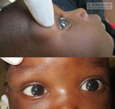
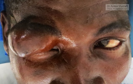
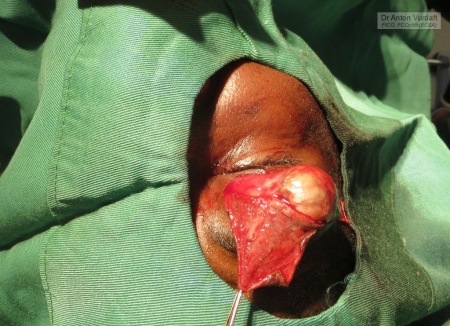
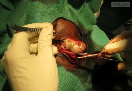
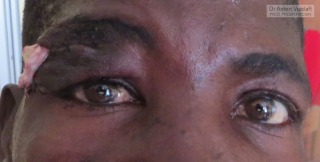
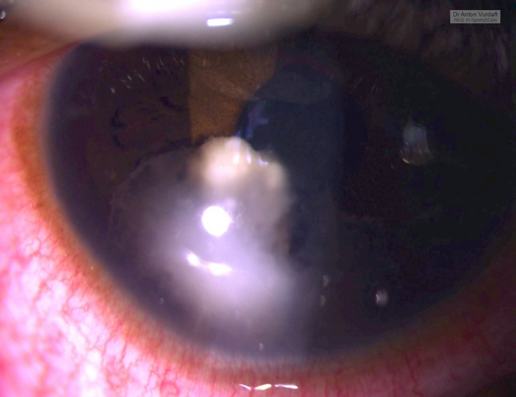
In EnglishHerpes zoster ophthalmicus involves V1 cranial nerve (n.ophthalmicus). Here are three examples of herpes zoster in 2 young men and one 7yo girl. Both men are HIV-positive (this must be an AIDS-stage). HZO is definitely second quickest germ to penetrate and destroy the cornea. Pseudomonas has no chance in comparison to those two. HIV is certainly a boost for the fulminant clinic.

in Russian / ΠΏΠΎ-русскиГСрпСс зостСр c Π²ΠΎΠ²Π»Π΅Ρ‡Π΅Π½ΠΈΠ΅ΠΌ ΠΏΠ΅Ρ€Π²ΠΎΠΉ Π²Π΅Ρ‚Π²ΠΈ ΠΈΠ· ΠΌΠΎΠ΅ΠΉ африканской ΠΏΡ€Π°ΠΊΡ‚ΠΈΠΊΠΈ. Π‘Π»ΡƒΡ‡Π°ΠΉ Ρƒ Π΄Π²ΡƒΡ… взрослых ΠΌΡƒΠΆΡ‡ΠΈΠ½ ΠΈ Ρƒ Π΄Π΅Π²ΠΎΡ‡ΠΊΠΈ 7 Π»Π΅Ρ‚ (Π°ΠΊΡ‚ΠΈΠ²Π½Ρ‹ΠΉ Ρƒ ΠΌΡƒΠΆΡ‡ΠΈΠ½, ΠΈ Π² исходС Ρƒ Π΄Π΅Π²ΠΎΡ‡ΠΊΠΈ). Herpes zoster ophthalmicus – Π½Π°Π²Π΅Ρ€Π½ΠΎΠ΅ Π²Ρ‚ΠΎΡ€ΠΎΠΉ ΠΏΠΎ скорости Ρ€Π°Π·ΡŠΠ΅Π΄Π°Π½ΠΈΡ Ρ€ΠΎΠ³ΠΎΠ²ΠΈΡ†Ρ‹ послС Π³ΠΎΠ½ΠΎΠΊΠΎΠΊΠΊΠΎΠ²ΠΎΠΉ ΠΈΠ½Ρ„Π΅ΠΊΡ†ΠΈΠΈ. БинСгнойная ΠΏΠ°Π»ΠΎΡ‡ΠΊΠ° ΠΎΠΏΡ€Π΅Π΄Π΅Π»Ρ‘Π½Π½ΠΎ Π½Π΅Ρ€Π²Π½ΠΎ ΠΊΡƒΡ€ΠΈΡ‚ Π² сторонкС. ΠœΡƒΠΆΡ‡ΠΈΠ½Ρ‹ Π±Ρ‹Π»ΠΈ ΠΎΠ΄Π½ΠΎΠ·Π½Π°Ρ‡Π½ΠΎ Π’Π˜Π§-ΠΏΠΎΠ»ΠΎΠΆΠΈΡ‚Π΅Π»ΡŒΠ½Ρ‹, Π° это, ΠΊΠ°ΠΊ извСстно, ΠΎΠ·Π½Π°Ρ‡Π°Π΅Ρ‚ ΡΡ‚Π°Π΄ΠΈΡŽ Π‘ΠŸΠ˜Π”. И это ΠΊΠΎΠ½Π΅Ρ‡Π½ΠΎ ΠΎΠ΄Π½Π° ΠΈΠ· основных ΠΏΡ€ΠΈΡ‡ΠΈΠ½ для Ρ‚Π°ΠΊΠΎΠΉ яркой ΠΈ драматичСской клиничСской ΠΊΠ°Ρ€Ρ‚ΠΈΠ½Ρ‹.

Advanced herpes zoster ophthalmicus with corneal melt and lens/uvea prolapse. HIV positive patient.

Advanced herpes zoster ophthalmicus with corneal melt and lens/uvea prolapse. HIV positive patient.

Advanced herpes zoster ophthalmicus with resultant corneal leucoma in 7yo girl.

Advanced herpes zoster ophthalmicus with resultant corneal leucoma in 7yo girl.

Advanced herpes zoster ophthalmicus with corneal melt and iris prolapse. HIV positive patient.

Advanced herpes zoster ophthalmicus with corneal melt and iris prolapse. HIV positive patient. Failed conjunctival flap adherence depicted.

Anterior chamber and iris granuloma in newly diagnosed HIV+ kid

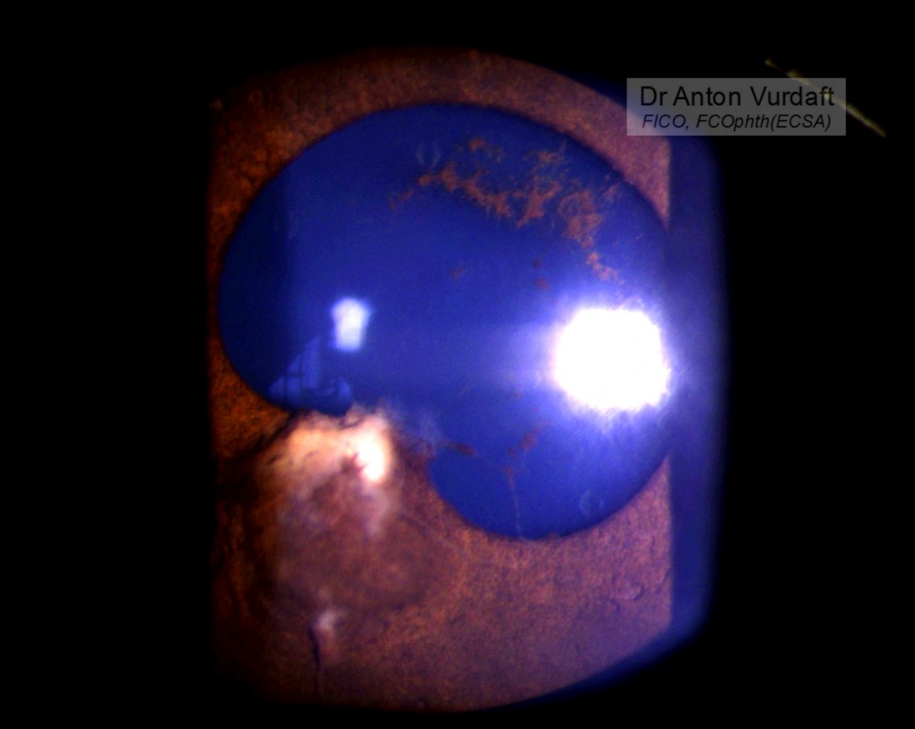
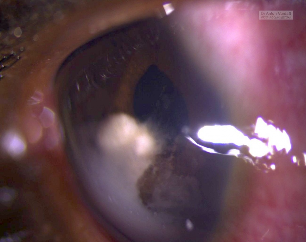
This was a case of Mai 2016. Eleven y.o. boy came to our outpatient department with the father complaining of “growth” in one of boy’s eyes, that was allegedly present already for 2 weeks. Visual acuity 6/9 and there were no pains except photophobia.Β  On examination – solid feathery cream-colored mass in the AC, adherant to the iris. Yellowish behind. No anterior chamber cells, no flare, absolutely quiet eye. Upon the HIV-test she was confirmed newly positive.

presumed aspergillosis iris-AC granuloma

presumed aspergillosis iris-AC granuloma

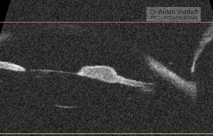
She was started on steroid and cycloplegic topical eye drops and NSAID tablets, and in 5 days the exudate was gone. But the iris cyst remained in place. There was no vitreous or retinal involvement, and the IOP was OK.

presumed aspergillosis iris-AC granuloma after tx, note torn posterior synechiae

presumed aspergillosis iris-AC granuloma after tx: UBM-scan

Differential diagnosis: intraocular medulloepithelioma (must be congenital, but looks similar, although originating mostly from the angle or ciliary body, not the iris), intraocular lymphoma (looks simmilar, but much more scary!), tuberculoma, leproma, nonpigment melanoma or primary iris tumors, toxoplasmosis gumma, metastases in the iris (lungs, intestine, kidneys), fungi!

Other useful resourcesΒ  to review iris cysts:

eyewiki.aao.org/Iris_Cysts
bjo.bmj.com/content/59/5/276.full.pdf
https://www.archivesofpathology.org/doi/pdf/10.5858/arpa.2010-0669-RS
http://www.ncbi.nlm.nih.gov/pmc/articles/PMC3872564/

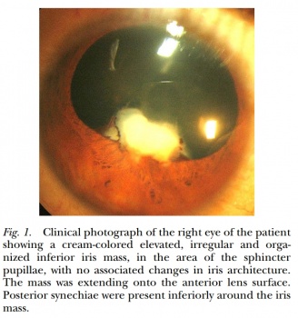
Literature search at that time in 2016 showed one paper from 2009 by Jain V with a similar case, Aspergillus iris granuloma: a case report with review of literature, claiming that to 2009 only three cases of aspergillosis of the anterior segment of the eye had been ever published. Here is a picture of their case:

Jain V, Aspergillus iris granuloma: a case report with review of literature. 2009

Our patient was left on the same topical treatment further in attempt to conquer the cyst and lost to follow-up. Unfortunately or fortunately, no iris-biopsy was performed that time, and therefore I had no microbiological evidence of fungi. One must also consider, that no natamycin was readily available topically in Western Province of Zambia that time, and in most of the times one must rely on antifungal activity of povidone-iodine eye drops (which are good and readily available hand made, by the way). One must strongly consider the lungs screening (CXR or CT) in immunocompromised patients, to exclude the primary source of hematogenous dissemination to
the brain or meninges. The immune status must be supported of course (f.e. HAART).

I thank my peer-colleagues from Terra-Ophthalmica for the kind help with the differential diagnosis and additional ideas for this case.
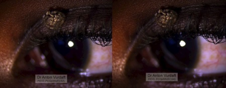
Peculiar golden eyelid papilloma

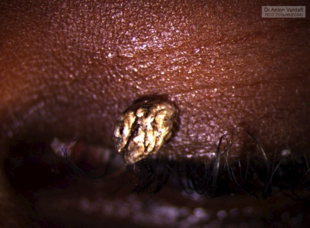
Golden upper eyelid papilloma

Golden upper eyelid papilloma

Stereo-pair image of Golden upper eyelid papilloma

Stereo-pair image of Golden upper eyelid papilloma

In EnglishA case of October 2016. A peculiar outlook of the upper eyelid papilloma in a 10yo girl. It was then immediately excised under topical anesthesia via shaving.


in Russian / ΠΏΠΎ-русски Золотая плоскоклСточная ΠΏΠ°ΠΏΠΈΠ»Π»ΠΎΠΌΠ° Π²Π΅Ρ€Ρ…Π½Π΅Π³ΠΎ Π²Π΅ΠΊΠ°. Π”Π΅Π²ΠΎΡ‡ΠΊΠ° 10 Π»Π΅Ρ‚. Π›Π΅Ρ‡Π΅Π½ΠΈΠ΅: прямоС иссСчСниС Π»Π΅Π·Π²ΠΈΠ΅ΠΌ Ρƒ основания ΠΏΠΎΠ΄ ΠΏΠΎΠ΄ΠΊΠΎΠΆΠ½ΠΎΠΉ анСстСзиСй (ΠΎΠ±Ρ€Π°Π±ΠΎΡ‚ΠΊΠ° области ΠΏΠΎΠ²ΠΈΠ΄ΠΎΠ½-ΠΉΠΎΠ΄ΠΎΠΌ, ΠΏΠΎΠ» ΠΊΡƒΠ±ΠΈΠΊΠ° ΠΈΠ»ΠΈ ΠΊΡƒΠ±ΠΈΠΊ Π»ΠΈΠ΄ΠΎΠΊΠ°ΠΈΠ½Π° ΠΏΠΎΠ΄ ΠΊΠΎΠΆΡƒ Π² этой области, Π»Π΅Π·Π²ΠΈΠ΅ Π±Ρ€ΠΈΡ‚Π²Ρ‹ ΠΈΠ»ΠΈ хирургичСский Π½ΠΎΠΆ β„–15, ΠΊΠΎΠΆΠ½Ρ‹ΠΉ ΠΏΠΈΠ½Ρ†Π΅Ρ‚, ΡΡ‚Π΅Ρ€ΠΈΠ»ΡŒΠ½Π°Ρ марля для купирования нСбольшого кровотСчСния, Π² повязкС Π½ΡƒΠΆΠ΄Ρ‹ Π½Π΅Ρ‚; выполняСтся Π² ΠΏΡ€ΠΎΡ†Π΅Π΄ΡƒΡ€Π½ΠΎΠΌ ΠΊΠ°Π±ΠΈΠ½Π΅Ρ‚Π΅).
Π‘Ρ‹Π» Π²Ρ‹Π½ΡƒΠΆΠ΄Π΅Π½ ΡΠ΄Π΅Π»Π°Ρ‚ΡŒ Ρ„ΠΎΡ‚ΠΎ, Ρ‚.ΠΊ. ΠΎΠ±ΠΈΠ΄Π½ΠΎ ΡƒΠ΄Π°Π»ΡΡ‚ΡŒ Ρ‚Π°ΠΊΠΎΠ΅ Π½Π΅ сфотографировав.