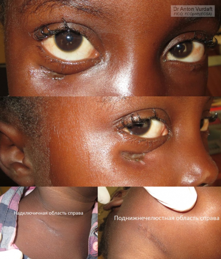
![]() EN: A case of August 2016. A 5yo girl with a fistula of right lower eyelid, discharging pus. The other striking feature was enlarged parotid lymph node at the same side, and a scar from the previous enlarged and drained by the general surgeon lymph nodes of the neck (that was some time back). The girl was afebrile, had unremarkable orbit and chest X-Ray. Unfortunately, blood analyses were not possible at the time in the Hospital. The case had remained a diagnostic dilemma, until a proper history was collected from a father. According to him, he had had a pulmonary TB in 2012 (the girl was born in 2011). The girl was started on standard TB treatment. Hereinafter the contact with them was lost.
EN: A case of August 2016. A 5yo girl with a fistula of right lower eyelid, discharging pus. The other striking feature was enlarged parotid lymph node at the same side, and a scar from the previous enlarged and drained by the general surgeon lymph nodes of the neck (that was some time back). The girl was afebrile, had unremarkable orbit and chest X-Ray. Unfortunately, blood analyses were not possible at the time in the Hospital. The case had remained a diagnostic dilemma, until a proper history was collected from a father. According to him, he had had a pulmonary TB in 2012 (the girl was born in 2011). The girl was started on standard TB treatment. Hereinafter the contact with them was lost.
Ideal management would have included Quantiferron TB test, PCR of the sputum, chest CT, orbit CT/MRI and lymph node biopsy.
Two remaining questions: would you biopsy the lymph node in this case? When and how would you repair the retracted and fistulized lower eye lid? (The soft tissues are adhered to the bone and are immobile in the area of fistula!).
Below are the links to the similar case reports from the literature. What is important, is that all of them: scrofuloderma, “cold” abscess, orbit rim osteomyelitis, periostitis, – are parts of the spectrum of tuberculous involvement of the orbit. “Scrofuloderma is a type of cutaneous tuberculosis affecting children and young adults. Also called tuberculosis colliquativa cutis, in this condition there is breakdown of skin overlying a tuberculous focus in the lymph node, bone, or joints.”
Orbital and adnexal tuberculosis: a case series from a South Indian population.
Tuberculous Orbital Abscess Associated with Thyroid Tuberculosis.
Tuberculous Periostitis of the Orbit: A Case Report. “The authors present a case of tuberculuos periostitis of the orbit which is extremely rare in nowadays. An 18-year-old man who had pulmonary and hepatic tuberculosis complained pain and erythematous swelling of periorbital region. Skull X-ray films showed multiple punched-out lesions on cranial vault. CT scan of the orbit revealed a homogenous mass on lateral orbital wall. The lesion was extended into the intracranial cavity with bony destruction and thickening of dura matter. The cold abscess of lower lid was drained by skin incision and Acid-fast bacilli was isolated from direct smear and culture.”
I extend my gratitude to the colleagues from Terra-Ophthalmica, whose help expedited the resolvation of diagnostic dilemma.
![]() RU: Случай августа 2016 г. Девочка 5 лет. Нижнее веко: углубление в нижнем веке с незаживающей хронической язвой у нижнего края орбиты, выделяющей небольшое количество гноя при надавливании на окружающие ткани. Язве 4 месяца. Температуры нет, общее самочувствие нормальное, состояние глаза нормальное (включая глазоподвижность). Веки тоже в остальном нормальны, но видна ретракция нижнего века (кожа в месте фистулы уплотнена, и спаяны с подлежащими тканями). Увеличен предушный лимфоузел, и нижнечелюстной узел справа. Орбиты по ультразвуку без какой-то грубой патологии. Со слов отца, “увеличенные лимфоузлы” имели место и справа на шее, и справа под нижней челюстью. Тот, что на шее – вскрывался в своё время хирургом. В то же время была лечена какими-то антибиотиками. Пазухи по рентгену без особенностей. Девочка не страдает иммунодефицитами.
RU: Случай августа 2016 г. Девочка 5 лет. Нижнее веко: углубление в нижнем веке с незаживающей хронической язвой у нижнего края орбиты, выделяющей небольшое количество гноя при надавливании на окружающие ткани. Язве 4 месяца. Температуры нет, общее самочувствие нормальное, состояние глаза нормальное (включая глазоподвижность). Веки тоже в остальном нормальны, но видна ретракция нижнего века (кожа в месте фистулы уплотнена, и спаяны с подлежащими тканями). Увеличен предушный лимфоузел, и нижнечелюстной узел справа. Орбиты по ультразвуку без какой-то грубой патологии. Со слов отца, “увеличенные лимфоузлы” имели место и справа на шее, и справа под нижней челюстью. Тот, что на шее – вскрывался в своё время хирургом. В то же время была лечена какими-то антибиотиками. Пазухи по рентгену без особенностей. Девочка не страдает иммунодефицитами.
Случай оставался диагностической дилеммой, до тех пор, пока отец на следующем приёме через 2 дня не поделился историей: он болел и лечил туберкулёз в 2012. Имел схожую лимфаденопатию на шее, завершившуюся рубцеванием, как у девочки. Девочка родилась в 2011.
До получения результатов рентгенографии лечил девочку внутривенными инъекциями цефтриаксона. Рентген орбиты и лёгких оказался без видимой патологии. Анализ крови в тот период был в госпитале недоступен.
Цитата из монографии проф. Устиновой Е.И. “Туберкулёз глаз и сходные с ним заболевания”: “Скрофулодерма – специфическое заболевание глубоких слоёв дермы и подкожной клетчатки, протекающее с образованием плотных узлов, последующим их размягчением и изъязвлением, образованием свищей. Скрофулодерма приводит к очень глубокому рубцеванию, анкилозу и несмыканию век. Туберкулёзный остеомиелит орбиты – почти всегда локализуется в её передней наружной половине, особенно в области нижне-наружного края. Характерно отсутствие болей, казеозный распад, абсцедирование с образованием свища. Фистулы в дальнейшем заживают распространённым, спаянным с костью рубцом, деформирующим веко”.
Пациентка была направлена к инфекционисту и начала лечение туберкулёза стандартной терапией. В дальнейшем контакт с ними был потерян. Остались неразрешёнными вопросы: 1) В каком объёме требовалась биопсия узла и ревизия орбиты? 2) На каком этапе и в каком объёме требовалась пластика нижнего века (фистула, ретракция, спаянные с костью ткани).
Литература с описанием схожих случаев представлена выше по ссылкам.
